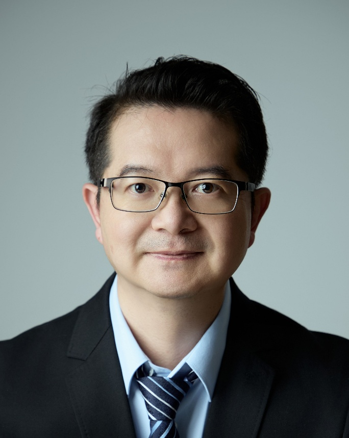
Faculty

Office address:Room 2513, Resources West Building, Peking University
Tel:010-62767155
mail:xipeng@pku.edu.cn
Homepage:www.xipenglab.com
Peng Xi
Professor
Boya Distinguished Professor
Dean, Dept. Biomedical Engineering
National Outstanding Young Scholar
Chief Scientist, Ministry of Science and Technology Key Program
Biography
Dr. Peng Xi is a full professor in College of Future Technology, Peking University, China. His research interest is on the development of optical super-resolution microscopy techniques. He has been awarded the National Outstanding Young Scholar. He has published over 80 scientific journal papers on peer-reviewed journals including Nature, Nature Methods, etc., and delivered over 30 keynote/invited talks on international conferences hosted by SPIE and OSA. He is a Senior member of Optica. He was the chair of OSA Imaging Optical Design Technical Group (2016-2019). He is on the editorial board of 5 SCI-indexed journals such as Light: Science and Applications, and Advanced Photonics.
Courses
n Biomedical Image Processing (UG, 3 credits)
n Biomedical Optics and Applications (UG, 3 credits)
Education
n BE 1998 Opto-electronic Engineering, Shanxi University
n Ph. D. 2003 Optical Engineering, Chinese Academy of Sciences (Supervisor: Prof. Changhe Zhou)
Experience
n 2003. 8 – 2004.12 Research Associate, Department of Electrical and Electronic Engineering, Hong Kong University of Science and Technology, Kowloon, Hong Kong (Advisor: Prof. Jianan Q. Qu)
n 2005. 2 – 2006.11 Research Associate, Bindley Bioscience Center, Purdue University, West Lafayette, IN, USA. (Advisor: Prof. J. Paul Robinson)
n 2006. 12 – 2007.12 Research Associate, Department of Chemistry, Michigan State University, East Lansing, MI, USA. (Advisor: Prof. Marcos Dantus)
n 2008. 1 – 2009.11 Associate Professor, School of Life Sciences and Biotechnology, Shanghai Jiao-Tong University, Shanghai, China
n 2009. 12 – 2021.1 Associate Professor, Department of Biomedical Engineering, College of Engineering, Peking University, Beijing, China
n 2021. 4 – now Professor, Department of Biomedical Engineering, College of Future Technology, Peking University, Beijing, China
Projects:
n 2021.1-2024.12 Principal Investigator, National Natural Science Foundation of China (No. 62025501)
Title: Super-resolution fluorescence microscopy (RMB 4,000,000)
n 2021.1-2024.12 Principal Investigator, National Natural Science Foundation of China (No. 62025501)
Title: Super-resolution fluorescence microscopy (RMB 4,000,000)
Representative publications:
(1) Y. Liu, Y. Lu*, X. Yang, X. Zheng, S. Wen, F. Wang, X. Vidal, T. Zhao, D. Liu, Z. Zhou, C. Ma, J. Zhou, J. Piper, P. Xi*, and D. Jin*, “Amplified stimulated emission in upconversion nanoparticles for super resolution nanoscopy,”Nature 543,229-233 (2017).
This paper reports the ultra-low power STED mechanism using the photo-avalanche effect of upconversion nanoparticle, with citation >800 times.
(2) K. Zhanghao*, W. Liu, M. Li, Z. Wu, X. Wang, X. Chen, C. Shan, H.n Wang, X. Chen, Q. Dai, P. Xi*, D. Jin*, High-dimensional super-resolution imaging reveals heterogeneity and dynamics of subcellular lipid membranes, Nature Communications, 11: 5890 (2020).
This work provides a new paradigm for multi-organelle interactome, leveraging the structured illumination, and the physical/chemical environmental differences in the organelles.
(3) X. Yang, Z. Yang, Z. Wu, Y. He, C. Shan, P. Chai, C. Ma, M. Tian, J. Teng, D. Jin, W. Yan, P. Das, J. Qu*, P. Xi*, “Mitochondrial dynamics quantitatively revealed by STED nanoscopy with an enhanced squaraine variant probe” Nature Communications 11:3699 (2020).
Development of a robust, squaraine variable mitochondria dye for long-term mitochondria STED imaging, with citation >120 times.
(4) R. Cao, Y. Li, X. Chen, X. Ge, M. Li, M. Guan, Y. Hou, Y. Fu, X. Xu, S. Jiang, B. Gao, P. Xi*, Open-3DSIM: an Open-source three-dimensional structured illumination microscopy reconstruction platform, Nature Methods 20, 1183-1186 (2023).
This paper offers the first open-source 3D-SIM reconstruction tool, to help boosting the development of 3D-SIM. We have also published the open-source 3D-SIM hardware on Advanced Photonics Nexus following this work.
(5) Chen X, Zhong S, Hou Y, Cao R, Wang W, Li D, Dai Q, Kim D, Xi P*. Superresolution structured illumination microscopy reconstruction algorithms: a review.Light: Science & Applications 12(1):172 (2023).
This work provides a review on the SIM reconstruction algorithms. ESI Highly Cited paper.
(6) K. Zhanghao*, X. Chen, M. Li, Y. Liu, W. Liu, S. Luo, X. Wang, K. Zhao, A. Lai, C. Shan, H. Xie, Y. Zhang, X. Li, Q. Dai*, P. Xi*, “Super-resolution Imaging of Fluorescent Dipoles by Polarized Structured Illumination Microscopy,” Nature Communications 10, 4694 (2019).
This work reports the polarization structured illumination microscopy. It has been highlighted by Nature Methods, and been cited >130 times.
(7) D. Jin*, P. Xi*, B. Wang, L. Zhang, J. Enderlein*, A. M. Oijen*, “Nanoparticles for super-resolution microscopy and single-molecule tracking", Nature Methods, 15, 415-423 (2018).
This review offers a comprehensive overview of the role of nanoparticles in super-resolution microscopy and single-molecule tracking, with citation >280 times.
(8) Xu, Xinzhu, Wenyi Wang, Liang Qiao, Yunzhe Fu, Xichuan Ge, Kun Zhao, Karl Zhanghao et al. "Ultra-high spatio-temporal resolution imaging with parallel acquisition-readout structured illumination microscopy (PAR-SIM)."Light:Science & Applications 13(1):125 (2024).
Improving the SIM imaging speed by 9.6-fold through active projecting the images onto the readout zone of sCMOS detector, enabling parallel acquisition and readout.
(9) Cao, R., Y. Li, Y. Zhou, M. Li, F. Lin, W. Wang, G. Zhang, G. Wang, B. Jin, W. Ren, Y. Sun, Z. Zhao, W. Zhang, J. Sun, Y. Hou, X. Xu, J. Hu, W. Shi, S. Fu, Q. Liang, Y. Lu, C. Li, Y. Zhao, Y. Li, D. Kuang, J. Wu, P. Fei, J. Qu and P. Xi (2025). "Dark-based optical sectioning assists background removal in fluorescence microscopy." Nature Methods (2025). https://doi.org/10.1038/s41592-025-02667-6
This paper reports the Dark sectioning algorithm which can boost the wide-field microscopy into confocal, single-photon to two-photon, 2D-SIM to 3D-SIM, and help more accurate segmentation for light-sheet and light-field microscopy.
(10) K. Zhanghao, M. Li*, X. Chen, W. Liu, T. Li, Y. Wang, F. Su, Z. Wu, C. Shan, J. Wu, Y. Zhang, J. Fu, P. Xi*, D. Jin*. "Fast Segmentation and Multiplexing Imaging of Organelles in Live Cells." Nature Communications 16, no. 1: 2769 (2025).
This work reports the segmentation and simultaneous imaging of 15 subcellular organelles. It has been highlighted by Nature Methods.
