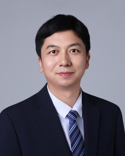
个人简介
主要研究方向为跨尺度纳米成像、大规模生物图像分析与建模,以及脑衰老机制研究。实验室致力于融合先进光学显微成像、PB级图像计算与人工智能技术,发展面向大体积生物组织的高分辨率、高通量三维成像及智能分析方法,突破跨尺度成像与大规模数据处理的技术瓶颈。基于海量成像数据,开展生物组织的数字化重建与多尺度计算建模,推动相关研究从结构观测向计算模拟与机制解析拓展,为探索脑衰老及神经退行性疾病的发生发展机制提供新方法。前期研究涵盖多模态自适应光学显微成像、PB级生物图像计算及大体积组织纳米成像等方向,以第一作者(含共同第一作者)或通讯作者身份在 Science、Nature Methods、Science Advances 等国际重要学术期刊发表论文,其中两篇论文分别入选 Science 和 Nature Methods 封面。
教育背景
2014-2019 卡内基梅隆大学,博士(计算生物学)
2008-2012 中国科学技术大学,学士(生物科学)
工作经历
2026.09-至今 北京大学未来技术学院 助理教授、研究员
2025.07-2026.08 加州大学伯克利分校 助理项目科学家
2019.07-2025.07 加州大学伯克利分校 博士后
2012.09-2014.04 杰克逊实验室 科研助理
代表性论文
* 表示共同第一作者;# 表示共同通讯作者
1. Fu, T.*, Liu, G.*, Milkie, D.E.*, Ruan, X.*, Görlitz, F., Shi, Y., Ferro, V., Divekar, N.S., Wang, W., York, H.M., Kilic, V., Mueller, M., Liang, Y., Daugird, T.A., Gacha-Garay, M.J., Larkin, K.A., Adikes, R.C., Harrison, N., Shirazinejad, C., Williams, S., Nourse, J.L., Sheu, S., Gao, L., Li, T., Mondal, C., Achour, K., Hercule, W., Stabley, D., Emmerich, K., Dong, P., Drubin, D., Liu, Z.J., Clapham, D., Mumm, J.S., Koyama, M., Killilea, A., Bravo-Cordero, J.J., Keene, C.D., Luo, L., Kirchhausen, T., Pathak, M.M., Arumugam, S., Nunez, J.K., Gao, R., Matus, D.Q., Martin, B.L., Swinburne, I.A., Betzig, E.#, Legant, W.R.#, Upadhyayula, S.# (2026). A multimodal adaptive optical microscope for in vivo imaging from molecules to organisms. Nature Methods, 23, 1184–1195 (Cover article)
2. Wang, W.*, Ruan, X.*, Liu, G.*, Milkie, D.E., Li, W., Betzig, E., Upadhyayula, S.#, Gao, R.# (2025). Mesoscale volumetric fluorescence imaging at nanoscale resolution by photochemical sectioning. Science, 390(6770), eadr9109 (Cover article; highlighted in Nature Biotechnology)
3. Ruan, X.#, Mueller, M., Liu, G., Görlitz, F., Fu, T., Milkie, D.E., Lillvis, J.L., Kuhn, A., Chong, J.G., Hong, J.L., Herr, C.Y.A., Hercule, W., Nienhaus, M., Killilea, A.N., Betzig, E.#, Upadhyayula, S.# (2024). Image processing tools for petabyte-scale light sheet microscopy data. Nature Methods, 21, 2342–2352
4. Liu, G.*, Ruan, X.*, Milkie, D.E., Görlitz, F., Mueller, M., Hercule, W., Killilea, A., Betzig, E.#, Upadhyayula, S.# (2023). Characterization, comparison, and optimization of lattice light sheets. Science Advances, 9(13), eade6623
5. Ruan, X.*, Johnson, G.R.*, Bierschenk, I., Nitschke, R., Boerries, M., Busch, H., Murphy, R.F. (2020). Image-derived models of cell organization changes during differentiation and drug treatments. Molecular Biology of the Cell, 31(7), 655–666
6. Ruan, X., Murphy, R.F. (2019). Evaluation of methods for generative modeling of cell and nuclear shape. Bioinformatics, 35(14), 2475–2485
7. Ruan, X., Wülfing, C., Murphy, R.F. (2017). Image-based spatiotemporal causality inference for protein signaling networks. Bioinformatics, 33(14), i217–i224 (ISMB 2017 proceedings)
8. Ambler, R.*, Ruan, X.*, Murphy, R.F.#, Wülfing, C.# (2017). Systems imaging of the immune synapse. The Immune Synapse: Methods and Protocols, 409–421
9. Roybal, K.T.*, Buck, T.E.*, Ruan, X.*, Cho, B.H., Clark, D.J., Ambler, R., Tunbridge, H.M., Zhang, J., Verkade, P., Wülfing, C.#, Murphy, R.F.# (2016). Computational spatiotemporal analysis identifies WAVE2 and cofilin as joint regulators of costimulation-mediated T cell actin dynamics. Science Signaling, 9(424), rs3