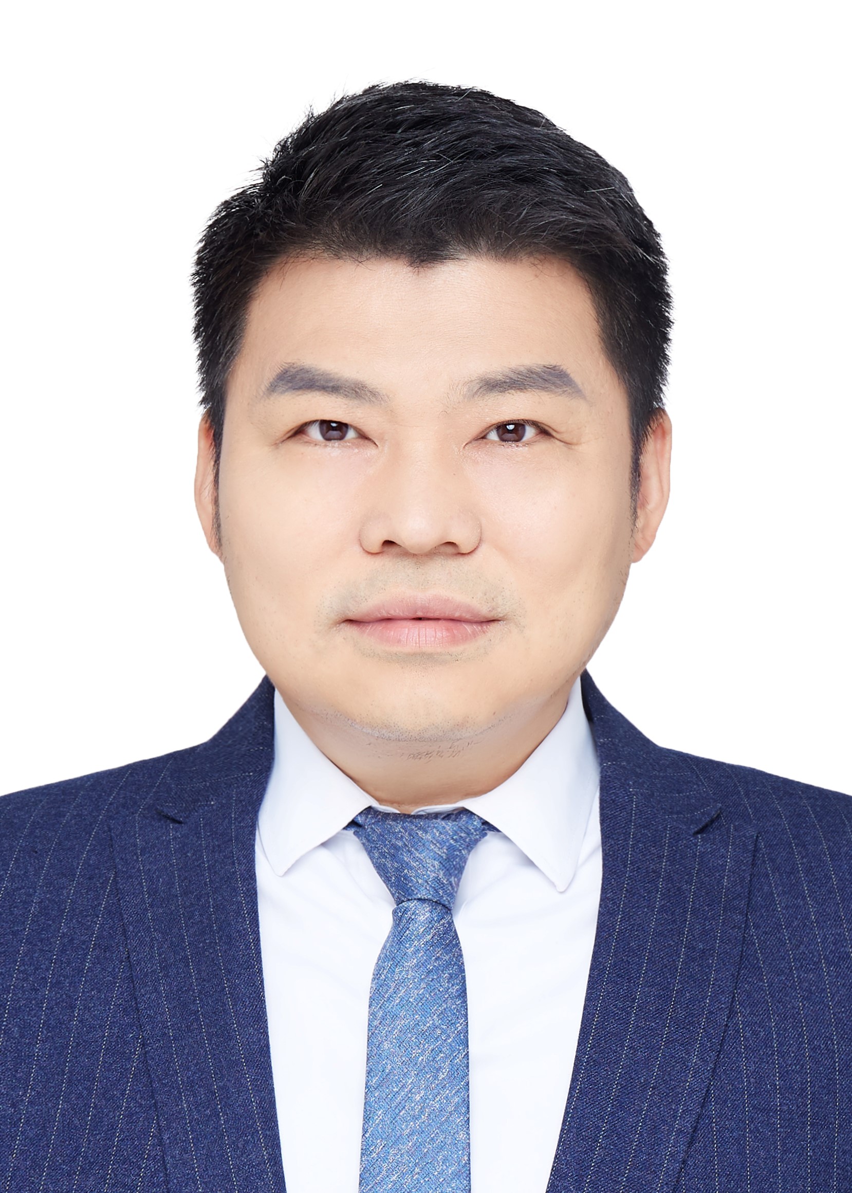
未来技术学院分子医学研究所博雅特聘教授、博士生导师
北京大学麦戈文脑研究所、北京智源人工智能研究院、北京市协同创新研究院兼职研究员
国家自然科学基金委杰出青年基金、优秀青年基金资助获得者
多模态跨尺度生物医学成像设施装置II 负责人
个人简介
主要研究方向是糖尿病发病过程中胰岛素分泌异常机制,发明了一系列的高时空分辨率生物医学成像的可视化手段,其主要科研发明及成就有:1)高分辨率微型化双光子显微镜,首次在自由行为小鼠上观察到单个神经突触的形态和功能变化,被2014年诺贝尔医学奖获得者Edvard Moser教授认为是“革命性”的新工具,工作入选了“2017年中国科学十大进展”等,也入选“Nature Methods: Method of the Year 2018”;2) 首次提出结构光显微镜在低信噪比时存在重建伪影的本质,发明海森结构光超分辨率显微镜,在光毒性最大降低了三个数量级下实现高保真度超分辨率成像,看见活细胞内线粒体内嵴以及胰岛素囊泡融合孔道,入选 “2018年中国光学十大进展”;3)活细胞双模态超分辨率显微镜,观察到细胞器互作全景图和新细胞器黑色液泡小体;4)提出活细胞超分辨率病理学的概念,利用活细胞超分辨率成像预测佩梅病临床疾病表型以及筛选出精准对症药物: 5)提出荧光信号具备稀疏和连续的通用先验,发明基于新数学原理的荧光计算超分辨率成像,与基于特定物理原理或者特殊荧光探针的传统超分辨率方法都不相同,实现活细胞成像中分辨率最高(60nm)、速度最快(564Hz)、成像时间最长(>1小时),获得“2021年度中国光学领域十大社会影响力事件(Light10)”和“首届全国颠覆性技术创新大赛”总决赛最高优胜奖。
发展的新成像技术推动了国内外许多合作者的生物医学基础研究工作,与来自国内外60个研究机构的近100个小组有科研合作,发表论文多篇;另一方面,将原创技术转化成为国内急需的高端显微镜产品,解决国内高端显微镜产品被国外厂商“卡脖子”的现状。作为创始人创立的两家专注生物医学成像技术的公司,商业化仪器销售额超过1亿元人民币,产品销往中国、欧洲和美国,使生物医学成像技术更加简单、易用。
视频版实验室简介:
https://mp.weixin.qq.com/s/fU-XKZs_op1fZBYwU2IlZg
所授课程
《高级生物物理》 (必修,2学分)
《生物荧光成像》 (选修,2学分)
《胰岛细胞生物学》 (选修,3学分)
教育背景
1990.9-1992.7 西安交通大学少年01班
1992.9-1995.7 西安交通大学生物工程与医学仪器系学士
1995.9-1998.7 华中理工大学自控系生物电子学硕士,师从康华光教授和瞿安连教授
1998.9-2001.4 华中科技大学生物医学工程博士,师从康华光教授和邹寿彬教授
工作简历
2001.4-2004.6 美国华盛顿大学生理和生物物理系 博士后,师从美国科学院院士Bertil Hille教授
2004.6-2010.6 中国科学院生物物理研究所 副研究员
2010.6-现在 北京大学分子医学研究所 研究员
2020.1-现在 北京大学分子医学研究所 博雅特聘教授
代表性论文
1. Zhao W, Zhao S, Li L, Huang X, Xing S, Zhang Y, Qiu G, Han Z, Shang Y, Sun D, Shan C, Wu R, Gu L, Zhang S, Chen R, Xiao J, Mo Y, Wang J, Ji W, Chen X, Ding B, Liu Y, Mao H, Song B, Tan J, Liu J, Li H*, Chen L*. Sparse deconvolution improves the resolution of live-cell super-resolution fluorescence microscopy, Nature Biotechnology. article in press.
2. Zong W, Wu R*, Chen S, Wu J, Wang H, Zhao Z, Chen G, Tu R, Wu D, Hu Y, Xu Y, Wang Y, Duan Z, Wu H, Zhang Y, Zhang Y, Wang A*, Chen L*, Cheng H. Miniature two-photon microscopy for enlarged field-of-view, multi-plane and long-term brain imaging, Nat Methods. 2021 Jan;18(1):46-49.
3. Zheng X, Duan R, Li L, Xing S, Ji H, Yan H, Gao K, Wang J, Wang J*, Chen L*. Live-cell superresolution pathology reveals different molecular mechanisms of Pelizaeus-Merzbacher disease, Science Bulletin, 30 December 2020; 65(24): 2061-2064.
4. Dong D, Huang X, Li L, Mao H, Mo Y, Zhang G, Zhang Z, Shen J, Liu W, Wu Z, Liu G, Liu Y, Yang H, Gong Q, Shi K*, Chen L*, Super-resolution fluorescence-assisted diffraction computational tomography reveals the three-dimensional landscape of the cellular organelle interactome, Light Sci Appl. 2020 Jan 28;9:11. doi: 10.1038/s41377-020-0249-4. eCollection 2020.
5. Zhao J, Zong W, Zhao Y, Gou D, Liang S, Shen J, Wu Y, Zheng X, Wu R, Wang X, Niu F, Wang A, Zhang Y, Xiong JW, Chen L*, Liu Y*. In Vivo imaging of β-cell function reveals glucose-mediated heterogeneity of β-cell functional development. Elife. 2019 Jan 29;8. pii: e41540. doi: 10.7554/eLife.41540.
Highlighted in the Nature Reviews Endocrinology (2019). https://doi.org/10.1038/s41574-019-0181-y;
6. Huang X, Fan J, Li L, Liu H, Wu R, Wu Y, Wei L, Mao H, Lal A, Xi P, Tang L, Zhang Y, Liu Y, Tan S*, Chen L*. Fast, long-term super-resolution imaging with Hessian structured illumination microscopy, Nat Biotech., 2018 Jun;36(5):451-459. doi: 10.1038/nbt.4115.
Highlighted in the Nature Methods (2018) https://doi.org/10.1038/s41592-018-0023-1. Hessian SIM has also been commented in the review in Nature Methods (2018) 15(12):1011-1019. doi:10.1038/s41592-018-0211-z as “the most sensitive super-resolution SIM microscope with photon dose one order less than other SIM modalities ”. 工作入选了“2018年中国光学十大进展”; Field-weighted citation impact (FWCI): 11.70, higher than 99% of similar works in the same field
https://www.youtube.com/watch?v=U9Om7DPxzd0&feature=youtu.be
7. Zong W, Wu R, Li M, Hu Y, Li Y, Li J, Rong H, Wu H, Xu Y, Lu Y, Jia H, Fan M, Zhou Z, Zhang Y*, Wang A*, Chen L*, Cheng H. Fast High-resolution Miniature Two-photon Microscopy for Brain Imaging in Freely-behaving Mice. Nat Methods. 2017 Jul;14(7):713-719.
An exclusive commentary ‘Miniaturized two-photon microscope: seeing clearer and deeper into the brain’ in Light: Science & Applications (2017) 6, e17104; doi:10.1038/lsa.2017.104. 工作入选了“2017年中国科学十大进展”,“2017年中国十大医学科技新闻”和“2017年中国生命科学十大进展”; “Nature Methods: Method of the Year 2018”; FWCI: 11.61, higher than 99% of similar works in the same field
https://www.youtube.com/watch?v=AED0SJVAlp0
8. Yuan T, Liu L, Zhang Y, Wei L, Zhao S, Zheng X, Huang X, Boulanger J, Gueudry C, Lu J, Xie L, Du W, Zong W, Yang L, Salamero J, Liu Y*, Chen L*. Diacylglycerol Guides the Hopping of Clathrin-Coated Pits along Microtubules for Exo-Endocytosis Coupling. Dev Cell. 2015 Oct 12;35(1):120-30.
Previewed in the same issue of Dev Cell, highlighted in the F1000Prime as NEW FINDING and INTERESTING HYPOTHESIS and also highlighted on the American Society of Cell Biology (ASCB) website.
9. Zong W, Zhao J, Chen X, Lin Y, Ren H, Zhang Y, Fan M, Zhou Z, Cheng H, Sun Y*, Chen L*. Large-field high-resolution two-photon digital scanned light-sheet microscopy. Cell Res. 2015 Feb;25(2):254-7.

